Researchers at Johns Hopkins University have created tiny EEG caps for brain organoids. The team was inspired by full size EEG caps that are used to measure brain activity in human patients. Previously, the Hopkins researchers were forced to use flat electrode arrays that were originally designed to take recordings from cell monolayers, but applying a flat surface to a round organoid only results in measurements from a handful of cells that make full contact. This latest technology consists of a tiny wrap-around EEG cap for the organoids, and consists of polymer-coated electrodes that are integrated into self-folding polymer leaflets. The device should lead to better data and more insights from brain organoids.
Organoids have been around for more than a decade, and offer enormous potential in providing new insights into human disease and in discovering new treatments. They also have huge benefits in reducing the number of experimental animals used in research labs while providing more human-specific data, as long as they are made with human cells. However, the tiny structures are, well… tiny, and they are also three dimensional, meaning that conventional cell culture equipment is not always best suited for interacting and assessing them. Most cell culture takes place in two dimensions with flat cell monolayers, which can vary wildly from organoids in their behavior and practical requirements in terms of conducting experiments.
 A brain organoid, shown in green, encapsulated in an electrodes device, depicted in blue.
Image credit: Qi Huang, Gayatri Pahapale, Gracias lab, Johns Hopkins University
Brain organoids, also sometimes called “mini-brains”, are particularly exciting, as they can offer insights into the human brain. “This provides an important tool to understand the development and workings of the human brain,” said David Gracias, a researcher involved in the study. “Creating micro-instrumentation for mini-organs is a challenge, but this invention is fundamental to new research.”
The Hopkins team behind this latest technology wanted to obtain more meaningful and accurate measurements from their brain organoids, and the poorly specific design of flat EEG equipment served as their motivation. A flat surface will only contact a small number of cells on the organoid surface. “We want to get information from as many cells as possible in the brain, so we know the state of the cells, how they communicate and their spatiotemporal electrical patterns,” said Gracias.
The result is a wrap-around EEG cap for individual brain organoids that allows 3D recording of many neurons within the organoid. “If you record from a flat plane, you only get recordings from the bottom of a 3D organoid sphere. However, the organoid is not just a homogeneous sphere,” said Qi Huang, another researcher involved in the study. “There are neuron cells that communicate with each other that’s why we need a spatial-temporal mapping of it.”
Study in journal Science Advances: Shell microelectrode arrays (MEAs) for brain organoids
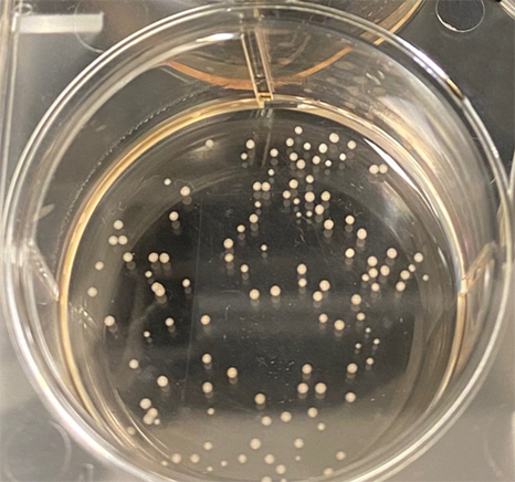
Top image: Organoids in a Petri dish. Image credit: Carolina Romero, Center for Alternative to Animal Testing, Bloomberg School of Public Health
